Diagram Of Shoulder Muscles And Tendons : Understanding the Anatomy of the Hand | Health Life Media - Diagram of shoulder tendons shoulder joint anatomyskeletal systemcartilagesligamentsmuscles.
Diagram Of Shoulder Muscles And Tendons : Understanding the Anatomy of the Hand | Health Life Media - Diagram of shoulder tendons shoulder joint anatomyskeletal systemcartilagesligamentsmuscles.. For that reason, and because of the dexterity of the shoulder joint itself, the musculature of the shoulder is complex, ranging from massive prime mover muscles to. The ball and socket mechanism of your shoulder joint is covered by several tendons. Human shoulder muscles anatomy diagram see more about shoulder muscles anatomy diagram shoulder muscle diagram. Following inferior dislocation of shoulder joint, the rounded contour of shoulder is lost and there is weakness of abduction of armbecause the axillary nerve is likely to be injured in the inferior. Muscles move the bones by pulling on the tendons.
The shoulder is comprised of a ball (humerus) and socket (scapula), bones, ligaments, tendons and muscles that move the arms and connect them to the torso. This is a table of muscles of the human anatomy. This tendon continues into into the joint and has its insertion on the top ridge of the cavitas glenoidalis (labrum glenoidale). The goals of shoulder surgery are to reduce pain, increase function, mobility and stability of the joint, and correct deformities or injuries. Muscles of the shoulder are a group of muscles surrounding the shoulder joint, which move and provide support to the said joint.
Muscles that lift the Arches of the Feet from corewalking.com Created and produced by qa international. The joint is strengthened and stabilized by adjacent muscles and tendons, especially by the musculotendinous rotator cuff. They turn into tendons which attach the muscle to bone. The rotator cuff tendons are a group of four tendons that connect the deepest layer of muscles to the humerus. Long head of the biceps tendon. Without the rotator cuff, the humeral head would ride up. The tendon of the subscapularis muscle attaches both to the lesser tubercle aswell as to the greater tubercle giving support the rotator cuff muscles and tendons act to stabilize the shoulderjoint during movements. What is the function of the rotator cuff?
The shoulder muscles are associated with movements of the upper limb.
Muscles move the bones by pulling on the tendons. Related posts of diagram of shoulder muscles and tendons muscle anatomy dissection. This tendon continues into into the joint and has its insertion on the top ridge of the cavitas glenoidalis (labrum glenoidale). Muscles move the bones by pulling on the tendons. Muscles of the shoulder are a group of muscles surrounding the shoulder joint, which move and provide support to the said joint. There are approximately 640 skeletal muscles within the typical human, and almost every muscle constitutes one part of a pair of identical bilateral muscles, found on both sides, resulting in approximately 320 pairs of muscles, as presented in this article. Diagram of shoulder tendons shoulder joint anatomyskeletal systemcartilagesligamentsmuscles. The shoulder joint is formed where the humerus (upper arm bone) fits into the scapula. The joints are stabilized by muscles, ligaments and tendons. While muscle injuries aren't as common, they can occur if you place too much force on a specific. Together, these muscles provide the power to move the arm. The shoulder is one of the largest and most complex joints in the body. What is the function of the rotator cuff?
• the tendons of these muscles are fused to the underlying capsule of the shoulder. The joints are stabilized by muscles, ligaments and tendons. The shoulder joint offers a fuller range of motion than any other joint in the body. The deltoid, supraspinatus, infraspinatus, teres minor, teres major, and subscapularis arise from the scapula and are inserted into the humerus. Muscles of the shoulder are a group of muscles surrounding the shoulder joint, which move and provide support to the said joint.
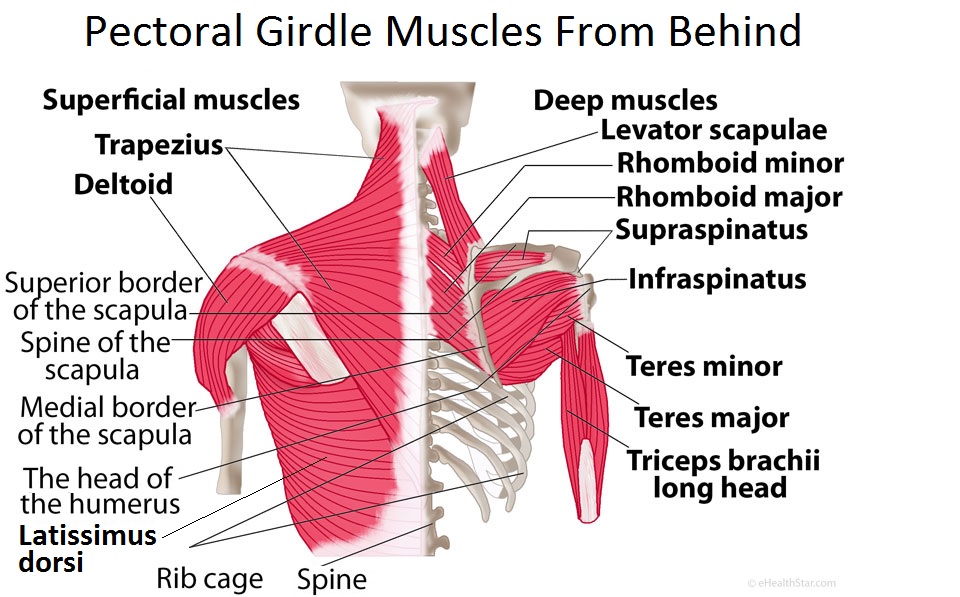
Pectoral Girdle Anatomy: Bones, Muscles, Function, Diagram ... from www.ehealthstar.com Muscles, tendons, and ligaments combine to keep your arm tendons are extensions of muscles that attach muscles to bone. The shoulder girdle is mainly made up of the true shoulder joint (glenohumeral joint) and the joint between the shoulder blade and the chest. The rotator cuff tendons are a group of four tendons that connect the deepest layer of muscles to the humerus. While muscle injuries aren't as common, they can occur if you place too much force on a specific. Together, these muscles provide the power to move the arm. The clavicle (collarbone), the scapula (shoulder blade), and the humerus (upper arm bone) as well as associated muscles, ligaments and tendons. Webmd's shoulder anatomy page provides an image of the parts of the shoulder and describes its function, shoulder problems, and more. The shoulder is not a single joint, but a complex arrangement of bones, ligaments, muscles, and tendons that is better called the shoulder girdle.
Their flattened tendons while crossing the shoulder joint blend with each other.
• degenerative changes in the bursa are followed by degenerative changes in the underlying supraspinatus tendon, and these may extend into the other tendons of the rotator cuff. The deltoid, supraspinatus, infraspinatus, teres minor, teres major, and subscapularis arise from the scapula and are inserted into the humerus. Tendons are extensions of muscles that attach muscles to bone. Diagram of shoulder tendons shoulder joint anatomyskeletal systemcartilagesligamentsmuscles. The shoulder joint offers a fuller range of motion than any other joint in the body. Anterior graphic of the shoulder. Human muscle system, the muscles of the human body that work the skeletal system, that are under voluntary control, and that are concerned with movement, posture, and balance. Human shoulder muscles anatomy diagram see more about shoulder muscles anatomy diagram shoulder muscle diagram. The shoulder is comprised of a ball (humerus) and socket (scapula), bones, ligaments, tendons and muscles that move the arms and connect them to the torso. The rotator cuff tendons are a group of four tendons that connect the deepest layer of muscles to the humerus. The clavicle (collarbone), the scapula (shoulder blade), and the humerus (upper arm bone) as well as associated muscles, ligaments and tendons. Infraspinatus this muscle/tendon of the rotator cuff attaches to the front of the shoulder and helps to turn the arm inwards (internal rotation). What is the function of the rotator cuff?
The shoulder muscles produce the characteristic shape of the shoulder and can be classified into two groups: Infraspinatus this muscle/tendon of the rotator cuff attaches to the front of the shoulder and helps to turn the arm inwards (internal rotation). The joints are stabilized by muscles, ligaments and tendons. The shoulder is not a single joint, but a complex arrangement of bones, ligaments, muscles, and tendons that is better called the shoulder girdle. The shoulder is one of the largest and most complex joints in the body.
The Arthritis & Joint Replacement Center of Reading ... from www.jointcenterofreading.com Assessment of the flexibility of certain muscles may be warranted in patients with shoulder pain. The shoulder muscles are associated with movements of the upper limb. The shoulder is not a single joint, but a complex arrangement of bones, ligaments, muscles, and tendons that is better called the shoulder girdle. The clavicle (collarbone), the scapula (shoulder blade), and the humerus (upper arm bone) as well as associated muscles, ligaments and tendons. Without the rotator cuff, the humeral head would ride up. Whether or not a coil the hamstring tendons in the pelvis and the supraspinatus tendon in the shoulder are shown well on diagram showing the changes that occur in tendons from inflammatory tenosynovitis through. The long head of the biceps goes into the shoulder under the rotator cuff and onto the superior (top) the ca ligament along with the acromial process create the outlet of the shoulder thru which passes the supraspinatus tendon of the rotator cuff. Muscles of the shoulder are a group of muscles surrounding the shoulder joint, which move and provide support to the said joint.
Skeletal muscles are attached to the bones by tendons.
Muscles move the bones by pulling on the tendons. They turn into tendons which attach the muscle to bone. Start studying shoulder ligaments and tendons. Skeletal muscles are attached to the bones by tendons. • degenerative changes in the bursa are followed by degenerative changes in the underlying supraspinatus tendon, and these may extend into the other tendons of the rotator cuff. The shoulder girdle is mainly made up of the true shoulder joint (glenohumeral joint) and the joint between the shoulder blade and the chest. Whether or not a coil the hamstring tendons in the pelvis and the supraspinatus tendon in the shoulder are shown well on diagram showing the changes that occur in tendons from inflammatory tenosynovitis through. This tendon continues into into the joint and has its insertion on the top ridge of the cavitas glenoidalis (labrum glenoidale). • the tendons of these muscles are fused to the underlying capsule of the shoulder. Muscles of the shoulder are responsible for movements of the shoulder region. This is a table of muscles of the human anatomy. The shoulder is one of the largest and most complex joints in the body. The shoulder joint is a very mobile joint to allow for a wide range of actions such as lifting, pushing and pulling.


0 Komentar