Loculated Pleural Effusion Ct Chest / Image-guided drainage of intrathoracic air and fluid ... - It refers to a fluid accumulation between the tissue layers lining the ct scan of the chest.
Loculated Pleural Effusion Ct Chest / Image-guided drainage of intrathoracic air and fluid ... - It refers to a fluid accumulation between the tissue layers lining the ct scan of the chest.. This is from increased pressure in the blood vessels or a low blood protein count. The pleura is a thin membrane that lines the inside of the chest wall and covers the lungs. Although pleural effusions are often easily identified on computed tomography (ct) the split pleura sign represents a rind of visceral and parietal pleural thickening surrounding a loculated effusion (figure 13). Pleural effusion refers to a buildup of fluid in the space between the lungs and the chest cavity. Terminology pleural effusion is commonly used as.
Figure 1 posteroanterior chest radiograph showing hypoinflation, cardiomegaly, indistinct vascular markings, and large bilateral pleural effusions. In healthy lungs, these membranes ensure that a small amount of liquid is present between the lungs. Pleural disease vs ____ disease 3. Prominent main pulmonary artery measuring 3.3 cm in diameter, which can be seen with pulmonary arterial hypertension. Clinical manifestations include chest pain, cough, and dyspnea.
Pleural Effusion Imaging: Overview, Radiography, Computed ... from img.medscapestatic.com Pleural effusions are abnormal accumulations of fluid within the pleural space. What is the key dx test for pleural effusion*. Recognize pleural calcification on a chest radiograph or ct and suggest the likely diagnosis of asbestos exposure (bilateral involvement) or old tuberculosis or trauma (unilateral involvement). Clinical manifestations include chest pain, cough, and dyspnea. The pleura is a thin membrane that lines the surface of your lungs and the inside of your chest wall. Prominent main pulmonary artery measuring 3.3 cm in diameter, which can be seen with pulmonary arterial hypertension. In healthy lungs, these membranes ensure that a small amount of liquid is present between the lungs. Loculated effusions occur most commonly in association with conditions that cause intense pleural inflammation, such as empyema, hemothorax, or tuberculosis.
It refers to a fluid accumulation between the tissue layers lining the ct scan of the chest.
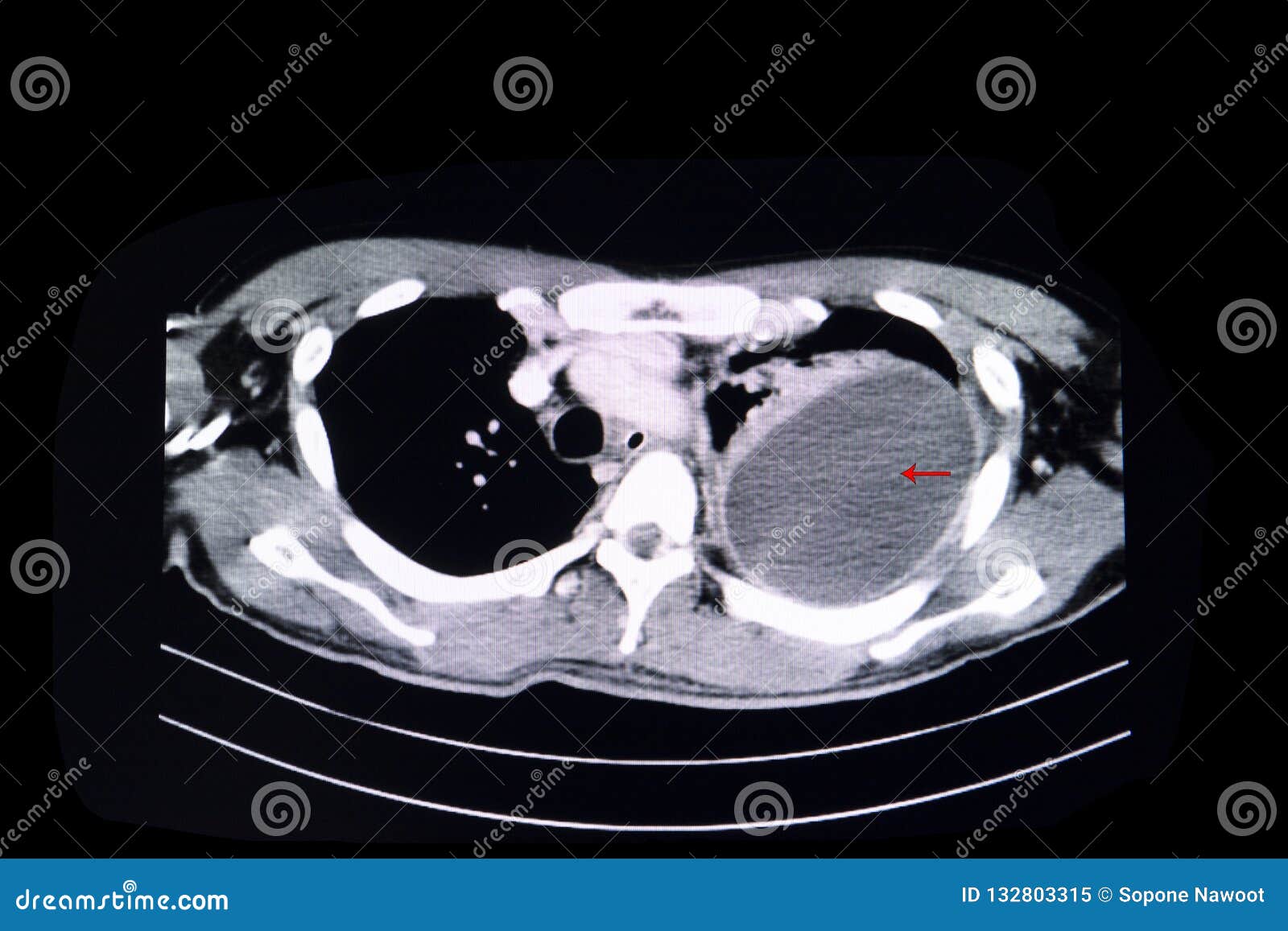
Transudative pleural effusion is caused by fluid leaking into the pleural space. The term pleura is generally meant to encompass the parietal pleura (lining the inner surface of the chest wall, including the diaphragmatic pleura and the cervical pleura also called dome of pleura or pleural. Bilateral, left greater than right, pleural effusions with adjacent atelectasis and collapse versus consolidation of the left lower lobe. Pleural thickening caused by tuberculosis can mimic a loculated pleural effusion is the major radiographic hallmark of parapneumonic effusion or empyema (see fig. Pleural disease vs ____ disease 3. Recognize the typical chest radiographic appearances of pleural effusion, given differences in. The pleura is a thin membrane that lines the inside of the chest wall and covers the lungs. Detection of pleural effusions on supine radiographs chest. Chest ct in pleural effusions 1. Ct scan of the chest of a patient with large loculated pleural effusion in his left thoracic cavity. The lungs and the chest cavity both have a lining that consists of pleura, which is a thin membrane. A pleural effusion is a buildup of fluid between the layers of tissue that line the lungs and chest cavity. A ct scan showing nodular, circumfrential pleural thickening and calcified pleural plaques in a patient who.
Terminology pleural effusion is commonly used as. What kind of effusions 4. Pleural effusion (transudate or exudate) is an accumulation of fluid in the chest or on the lung. Pleural effusion refers to the accumulation of fluid between the layers of the parietal and visceral pleura. Chest ct in pleural effusions 1.
Investigation of a unilateral pleural effusion in adults ... from thorax.bmj.com The plain chest radiographic features of pleural effusion are usually characteristic. Other imaging tests, such as ct scan, may be ordered to further identify the possible cause and the. Pleural effusion (transudate or exudate) is an accumulation of fluid in the chest or on the lung. The pleura is a thin membrane that lines the surface of your lungs and the inside of your chest wall. Transudative pleural effusion is caused by fluid leaking into the pleural space. Recognize the typical chest radiographic appearances of pleural effusion, given differences in. Chest ct in pleural effusions 1. More than one half of these massive pleural effusions are caused by malignancy;
Common causes of this condition include infection, malignancy, autoimmune disorders, or volume overload.
Clinical manifestations include chest pain, cough, and dyspnea. Ct scan of the chest of a patient with large loculated pleural effusion in his left thoracic cavity. The pleura is a thin membrane that lines the inside of the chest wall and covers the lungs. Figure 1 posteroanterior chest radiograph showing hypoinflation, cardiomegaly, indistinct vascular markings, and large bilateral pleural effusions. Recognize pleural calcification on a chest radiograph or ct and suggest the likely diagnosis of asbestos exposure (bilateral involvement) or old tuberculosis or trauma (unilateral involvement). Other imaging tests, such as ct scan, may be ordered to further identify the possible cause and the. The plain chest radiographic features of pleural effusion are usually characteristic. The pleura are thin membranes that line the lungs and the inside of the chest cavity and act to lubricate and facilitate breathing. Pleural thickening caused by tuberculosis can mimic a loculated pleural effusion is the major radiographic hallmark of parapneumonic effusion or empyema (see fig. The pleura is a thin membrane that lines the surface of your lungs and the inside of your chest wall. A ct scan showing nodular, circumfrential pleural thickening and calcified pleural plaques in a patient who. Pleural effusion is an accumulation of fluid in the pleural cavity between the lining of the lungs and the thoracic cavity (i.e., the visceral and parietal pleurae). Learn more from webmd about different types of pleural effusions,including symptoms, causes, and treatments.
Terminology pleural effusion is commonly used as. The pleura is a thin membrane that lines the inside of the chest wall and covers the lungs. Clinical manifestations include chest pain, cough, and dyspnea. Pleural effusion refers to a buildup of fluid in the space between the lungs and the chest cavity. Blood tests to check functioning of the kidneys and the liver.
Loculated pleural effusion stock image. Image of computer ... from thumbs.dreamstime.com Pleural effusion (transudate or exudate) is an accumulation of fluid in the chest or on the lung. Recognize pleural calcification on a chest radiograph or ct and suggest the likely diagnosis of asbestos exposure (bilateral involvement) or old tuberculosis or trauma (unilateral involvement). They may result from a variety of pathological processes which overwhelm the pleura's ability to reabsorb fluid. The pleura are thin membranes that line the lungs and the inside of the chest cavity and act to lubricate and facilitate breathing. Terminology pleural effusion is commonly used as. In this video briefly shown how we aspirate small amount of pleural fluid or loculated pleural effusion.for more videos please subscribe the channel.if you. Other causes are complicated parapneumonic effusion. Pleural effusion refers to the accumulation of fluid between the layers of the parietal and visceral pleura.
Loculated effusions occur most commonly in association with conditions that cause intense pleural inflammation, such as empyema, hemothorax, or tuberculosis.
It refers to a fluid accumulation between the tissue layers lining the ct scan of the chest. The term pleura is generally meant to encompass the parietal pleura (lining the inner surface of the chest wall, including the diaphragmatic pleura and the cervical pleura also called dome of pleura or pleural. Other causes are complicated parapneumonic effusion. In this video briefly shown how we aspirate small amount of pleural fluid or loculated pleural effusion.for more videos please subscribe the channel.if you. Other imaging tests, such as ct scan, may be ordered to further identify the possible cause and the. Learn more from webmd about different types of pleural effusions,including symptoms, causes, and treatments. Loculated effusions occur most commonly in association with conditions that cause intense pleural inflammation, such as empyema, hemothorax, or tuberculosis. Pleural disease vs ____ disease 3. Although pleural effusions are often easily identified on computed tomography (ct) the split pleura sign represents a rind of visceral and parietal pleural thickening surrounding a loculated effusion (figure 13). Recognize the typical chest radiographic appearances of pleural effusion, given differences in. The effusion may cause you to become breathless. Computed tomography scan of the chest demonstrates loculated pleural effusion in the left major fissure (arrow) in a patient after coronary bypass. They may result from a variety of pathological processes which overwhelm the pleura's ability to reabsorb fluid.
What kind of effusions 4 loculated pleural effusion ct. A pleural effusion is a collection of fluid next to the lung.


0 Komentar